Technology

Technology
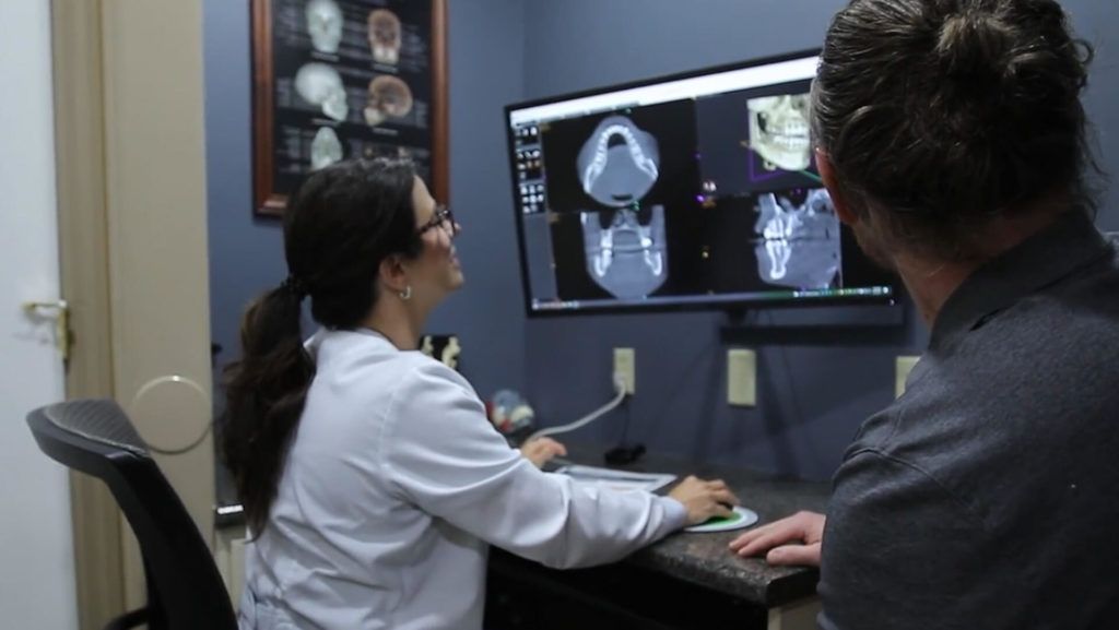
Technology in the dental field is ever-changing and Dr. Labritz and the Sorriso Dental team enjoy learning how to integrate state-of-the-art equipment into their practice. Utilizing the latest advancements in dentistry allows Dr. Labritz and the Sorriso Dental team to treat patients with less pain and in fewer visits which equals less time in the dental chair and a more comfortable experience from start to finish.
Dr. Labritz and the Sorriso Dental team and their team spend many hours each year training and taking advanced courses to ensure they deliver the best care, safely. Employing the latest technology means consistent results, so you’ll know what to expect at each visit. You’ll find that modern dental care with Sorriso Dental leaves you smiling.
At Sorriso Dental, you’ll discover dental care at its highest level; technology, advanced training, and compassionate service blended perfectly together.
High magnification
4k microscope
The Motic High Magnification Microscope is a revolutionary instrument in modern dental care and a significant tool for diagnosing periodontal disease. It functions by enhancing the contrast in transparent and colorless specimens, enabling the visualization of internal structures without the need for staining or tagging. By leveraging this technology, our dentists can promptly identify the types of bacteria present in a patient’s mouth, identifying high-risk bacteria much earlier than traditional methods. The Motic High Magnification Microscope is particularly remarkable because it not only analyzes bacteria but can also detect the presence of an infection within minutes. Its capabilities extend to identifying and assessing bacteria motility (movement), allowing for a deeper understanding of the microbial environment in the patient's mouth.


Dr. Caroline Labritz and her team at Sorriso Dental have effectively incorporated the use of the Motic High Magnification Microscope in their practice to combat periodontal disease. The comprehensive preventative care exam they conduct involves using a digital dental microscope to inspect patients' oral health minutely.
On microscopic slides, they assess the levels of bacteria, white blood cells, and some red blood cells, looking for low motility and low bacteria count, specifically rods and spirochetes. These are known to be the primary culprits behind gum and bone destruction. When spirochetes are discovered, a bactericidal agent is applied to irrigate the gums before any invasive dental treatment, thus preventing bacteremia.
Their approach extends to monitoring the bacteria under a microscope during periodontal wellness appointments and tailoring toothpaste and mouthwash to the bacteria found in the periodontal pocket. Through real-time visual images of the bacteria, patients become more compliant with the treatment, contributing significantly to their journey towards oral health recovery.
Intra-oral Camera
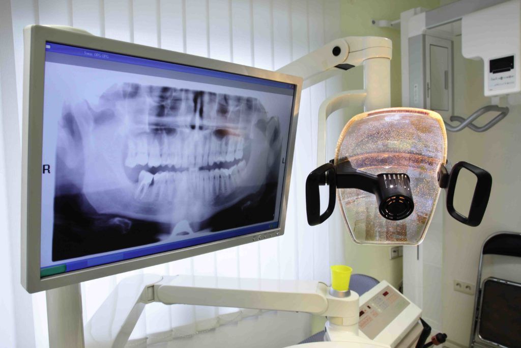
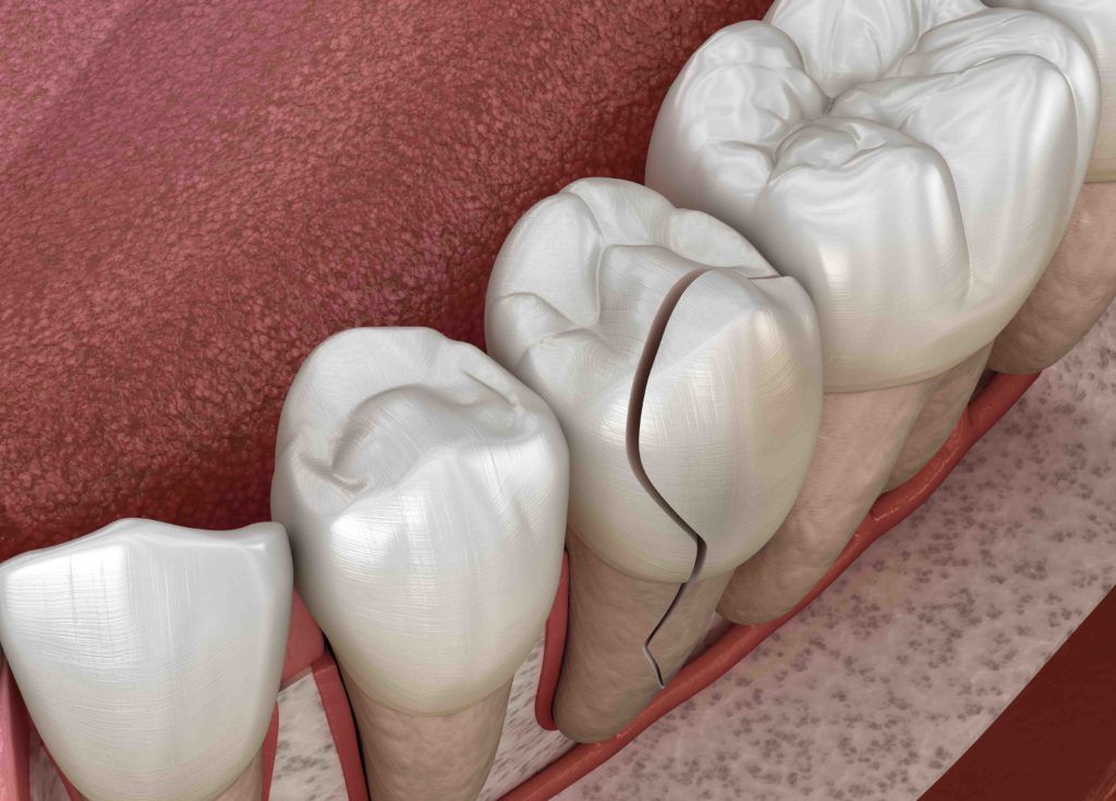
A picture explains what words never can, especially when it comes to your teeth. An intra-oral camera can take pictures with such detail, it can show cracks and stress marks in a tooth that can’t be seen with the naked eye.
Intra-oral imaging gives you a detailed view of your mouth in pictures and videos. Words come to life so you can see what we see.
Putting specific problem areas onto a computer screen lets us see with a clarity and detail not possible with a traditional mouth mirror. Specialized hand-held wands quickly capture the details of your teeth and gums. In fact, even small cracks that often lead to broken teeth appear vividly, with the click of a button. We can explain the need for treatment more clearly, by showing you what we see and with greater detail.
As an added benefit, dental insurance benefits frequently pay easier with the visual proof of a captured image in our records.